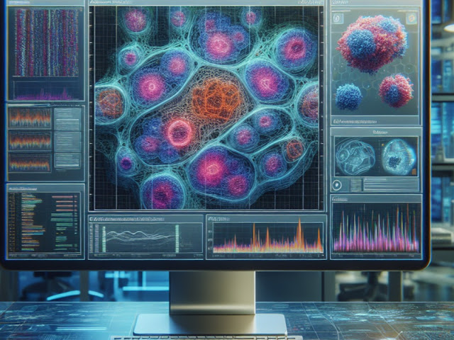
The Perelman School of Medicine at the University of Pennsylvania has revealed a groundbreaking artificial intelligence (AI) tool called iStar that could revolutionize pathology research. This remarkable tool, detailed in a recent publication in Nature Biotechnology, offers unprecedented insights into tissue microenvironments and gene activity, accelerating the understanding of diseases and potential treatments.
iStar can capture intricate details while also providing a broader structural context of tissue samples. It can detect "tertiary lymphoid structures," which are linked to anti-tumor responses and a patient's chances of survival and positive outcomes from immunotherapy.
One impressive feature of iStar is its speed. The tool's rapid processing capabilities enable researchers to reconstruct vast amounts of spatial data in a short period of time. This increased efficiency will have a profound impact on the scale and scope of pathology research, enabling the analysis of more tissue samples and a deeper understanding of tissue microenvironments.
By using AI algorithms, iStar provides insights into gene activity within cells, a crucial aspect of understanding diseases at the molecular level. This integration of AI and high-resolution imaging allows researchers to uncover hidden patterns and correlations that hold significant implications for diagnosis, treatment, and personalized medicine.
The potential applications of iStar are diverse and extensive. Researchers aim to leverage this groundbreaking technology to gain a comprehensive understanding of tissue microenvironments in various diseases, including cancer, autoimmune disorders, and infectious diseases. By unlocking new insights into the complexities of these diseases, iStar has the potential to pave the way for targeted therapies and more effective treatment strategies.
The development of iStar is a significant milestone in pathology research. By combining state-of-the-art imaging technology and AI algorithms, iStar provides a comprehensive view of tissue architecture, allowing researchers to explore the intricate details of cellular structures while also examining the broader context. This integration enhances our understanding of disease progression and response to treatment.
The impact of iStar extends beyond research, as it has the potential to improve diagnostics and prognostics. By enabling a more precise assessment of tissue microenvironments, this technology could lead to more accurate diagnoses, personalized treatment plans, and improved patient outcomes. Furthermore, iStar could facilitate the development of new biomarkers and therapeutic targets, ushering in a new era of precision medicine.
As the field of pathology research continues to evolve, iStar represents a significant leap forward. Combining AI, high-resolution imaging, and rapid data processing capabilities, this revolutionary tool has the potential to reshape our understanding of diseases and transform the approach to diagnostics and treatment. With iStar, researchers now have an unprecedented opportunity to unlock the mysteries of tissue microenvironments, paving the way for groundbreaking discoveries and advances in patient care.
The introduction of iStar marks a groundbreaking development in the field of pathology research. This revolutionary AI tool combines high-resolution imaging with rapid data processing capabilities to provide unparalleled insights into tissue microenvironments and gene activity. With its potential to revolutionize diagnostics, treatment strategies, and personalized medicine, iStar is poised to transform the landscape of pathology research and improve patient outcomes. The future of pathology research has arrived, and it looks brighter than ever with iStar paving the way for new discoveries and advancements in healthcare.


0 Comments